
Brain Anatomy: A Comprehensive Overview
Brain anatomy resources, often found as PDF documents, detail the complex structures of the human nervous system, including the cerebrum, brainstem, and cerebellum.
These diagram-rich guides, adapted from texts like Marieb and Hoehn’s Human Anatomy & Physiology, define key parts for labeling and understanding.
They cover the gross anatomy, cellular components – neurons and glial cells – and clinical relevance for diagnosis, weighing in at 1.3-1.5 Kg for adults.
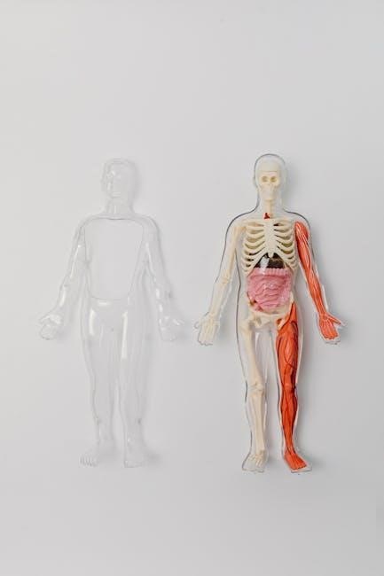
Brain anatomy, a cornerstone of neurological study, is frequently explored through detailed PDF resources offering comprehensive visual and textual guides. These materials present the intricate organization of the central nervous system, encompassing the brain and spinal cord, and the peripheral nervous system.
Understanding brain anatomy necessitates recognizing its division into major parts: the cerebrum, responsible for higher-level functions; the brainstem, connecting to the spinal cord; and the cerebellum, crucial for coordination. Many PDFs utilize embryonic schemes to illustrate developmental origins and structural relationships.
These resources often feature labeled diagrams, aiding in the identification of structures like the cerebellum, corpus callosum, and dentate nucleus. The adult human brain, averaging 1.3-1.5 Kg, showcases remarkable complexity. PDF documents frequently draw upon established texts like Marieb and Hoehn’s Human Anatomy & Physiology, providing a standardized and reliable foundation for learning. They also detail the roles of oligodendrocytes and Schwann cells.
II. Gross Anatomy of the Brain
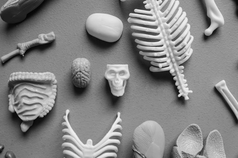
PDF resources dedicated to brain anatomy emphasize the macroscopic structures visible to the naked eye – the gross anatomy. These materials typically begin with a broad overview, highlighting the three primary divisions: the cerebrum, the brainstem, and the cerebellum. Detailed diagrams within these PDFs illustrate the convoluted surfaces and relative sizes of each component.
The cerebrum, the largest part, is often depicted with its characteristic gyri and sulci. The brainstem, acting as a stalk, connects the cerebrum to the spinal cord, and its structure is clearly outlined. The cerebellum, known for its rippled appearance, is also thoroughly illustrated.
These PDF guides, frequently adapted from texts like Marieb and Hoehn’s Human Anatomy & Physiology, also showcase the olfactory bulbs, though diminutive. Understanding these major parts is foundational, as they communicate with different areas of the brain and cerebellar cortex, as detailed in labeled anatomical charts.
A. Cerebrum: Structure and Function
PDF guides on brain anatomy dedicate significant attention to the cerebrum, the largest and most complex part of the human brain. These resources detail its structure, emphasizing the prominent gyri (ridges) and sulci (grooves) that increase surface area. Diagrams within these PDFs often showcase the longitudinal fissure dividing the cerebrum into left and right hemispheres.
Functionally, the cerebrum is responsible for higher-level cognitive processes. PDF materials explain how the cerebrum integrates sensory information and initiates voluntary motor responses. Resources adapted from texts like Marieb and Hoehn’s Human Anatomy & Physiology often correlate specific cerebral areas with functions like reasoning, memory, and language.
The corpus callosum, a crucial structure connecting the hemispheres, is also highlighted in these PDFs. Understanding the cerebrum’s intricate structure is vital, given its weight contributes significantly to the brain’s overall 1.3-1.5 Kg mass.
B. Brainstem: Connecting the Brain to the Spinal Cord
Brain anatomy PDF resources consistently emphasize the brainstem’s critical role as the vital link between the cerebrum and the spinal cord. Diagrams within these guides clearly illustrate its three main components: the midbrain, pons, and medulla oblongata. These PDFs detail how the brainstem’s structure facilitates the transmission of motor and sensory signals.
Functionally, the brainstem controls essential life-sustaining processes. PDF materials explain its involvement in regulating heart rate, breathing, and blood pressure. Resources, often adapted from comprehensive texts, highlight the cranial nerves originating from the brainstem and their associated functions.
The brainstem’s relatively small size – a distinct part of the brain’s 1.3-1.5 Kg mass – belies its immense importance. These PDFs underscore that damage to the brainstem can have devastating consequences, impacting fundamental physiological functions.
III. Lobes of the Cerebral Cortex
Brain anatomy PDF guides dedicate significant sections to the cerebral cortex and its four distinct lobes: frontal, parietal, temporal, and occipital. These resources utilize detailed diagrams to visually delineate each lobe’s boundaries and internal structures. The PDFs emphasize that understanding lobar functions is crucial for comprehending overall brain activity.
These materials, often adapted from sources like Human Anatomy & Physiology, explain how each lobe specializes in different cognitive and sensory processes. The frontal lobe, responsible for executive functions, receives extensive coverage. Similarly, the PDFs detail the parietal lobe’s role in sensory processing, the temporal lobe’s involvement in auditory and memory functions, and the occipital lobe’s specialization in visual processing.
These PDFs consistently highlight the interconnectedness of the lobes, noting that complex tasks often require the coordinated activity of multiple cortical areas within the brain’s 1.3-1.5 Kg mass.
A. Frontal Lobe: Executive Functions
Brain anatomy PDF resources extensively detail the frontal lobe, emphasizing its role in “executive functions.” These PDF guides, often featuring labeled diagrams, illustrate the lobe’s location and key internal structures. They explain how the frontal lobe, a significant part of the brain’s 1.3-1.5 Kg weight, governs higher-level cognitive processes.
These materials, adapted from texts like Marieb and Hoehn’s, clarify that executive functions encompass planning, decision-making, working memory, and impulse control. The PDFs highlight the frontal lobe’s connection to personality and social behavior. They also explain how damage to this area can result in significant behavioral and cognitive deficits.
Furthermore, these resources often include clinical case studies demonstrating the impact of frontal lobe dysfunction, reinforcing the importance of understanding its anatomy and function as detailed in the PDFs.
B. Parietal Lobe: Sensory Processing
Brain anatomy PDF documents dedicate significant attention to the parietal lobe, detailing its crucial role in sensory processing. These resources, often incorporating detailed diagrams, illustrate the lobe’s location and internal organization, contributing to the overall 1.3-1.5 Kg weight of the adult human brain.
PDF guides, frequently adapted from comprehensive texts like those by Marieb and Hoehn, explain how the parietal lobe integrates sensory information – including touch, temperature, pain, and spatial awareness. They clarify its involvement in navigation and understanding the body’s position in space.
These materials emphasize the parietal lobe’s connection to other brain regions, highlighting its role in complex cognitive functions. Clinical examples within the PDFs demonstrate the consequences of parietal lobe damage, such as sensory neglect or difficulties with spatial reasoning.
C. Temporal Lobe: Auditory and Memory Functions
Brain anatomy PDF resources extensively cover the temporal lobe, emphasizing its critical functions in auditory processing and memory formation. Detailed diagrams within these documents illustrate the lobe’s internal structures, contributing to the overall complexity of the 1.3-1.5 Kg adult human brain.
These PDF guides, often adapted from texts like Marieb and Hoehn’s Human Anatomy & Physiology, explain how the temporal lobe receives and interprets auditory information, enabling us to perceive and understand sounds. They also detail its role in long-term memory consolidation.
The materials highlight the temporal lobe’s connection to the limbic system, explaining its involvement in emotional memory. Clinical case studies within the PDFs demonstrate the effects of temporal lobe damage, such as amnesia or auditory agnosia, providing valuable diagnostic insights.
D. Occipital Lobe: Visual Processing
Brain anatomy PDF materials dedicate significant attention to the occipital lobe, detailing its primary role in visual processing. These resources, often derived from comprehensive texts like Marieb and Hoehn’s Human Anatomy & Physiology, feature detailed diagrams illustrating the lobe’s intricate structure within the 1.3-1.5 Kg adult human brain.
The PDFs explain how the occipital lobe receives visual information from the eyes and interprets it, enabling us to perceive shapes, colors, and motion. They delineate the visual pathways and cortical areas responsible for different aspects of vision.
Clinical examples within these documents showcase the consequences of occipital lobe damage, such as cortical blindness or visual agnosia, aiding in diagnostic understanding. The materials also explore the lobe’s connections to other brain regions, highlighting its contribution to spatial awareness and visual-motor coordination.
IV. Deep Brain Structures
Brain anatomy PDF resources extensively cover the deep brain structures, emphasizing their crucial roles despite being hidden beneath the cerebral cortex. These documents, often adapted from sources like Human Anatomy & Physiology, utilize detailed diagrams to illustrate the diencephalon – specifically the thalamus and hypothalamus – and the limbic system.
The PDFs explain how the thalamus acts as a relay station for sensory information, while the hypothalamus regulates vital functions like body temperature and hunger. They detail the limbic system’s involvement in emotion, motivation, and memory formation.
Furthermore, these materials explore the interconnectedness of these structures, demonstrating how they contribute to overall brain function within the 1.3-1.5 Kg human brain. Clinical correlations within the PDFs highlight the impact of damage to these areas, aiding in diagnostic comprehension.
A. Diencephalon: Thalamus and Hypothalamus
Brain anatomy PDF guides dedicate significant sections to the diencephalon, meticulously detailing the thalamus and hypothalamus. These resources, often derived from comprehensive texts, employ labeled diagrams to illustrate their location and intricate connections within the brain.
The PDFs explain the thalamus’s role as a crucial relay station, processing and transmitting sensory information to the cerebral cortex. They highlight its involvement in motor control, consciousness, and sleep. Simultaneously, they detail the hypothalamus’s function in maintaining homeostasis, regulating body temperature, hunger, and thirst.
These documents emphasize the hypothalamus’s connection to the pituitary gland, controlling hormonal release. Clinical notes within the PDFs often correlate damage to these structures with specific neurological deficits, within the context of the adult human brain weighing 1.3-1.5 Kg.

B. Limbic System: Emotion and Motivation
Brain anatomy PDF resources extensively cover the limbic system, illustrating its key structures – amygdala, hippocampus, and cingulate gyrus – through detailed diagrams. These guides, often adapted from sources like Human Anatomy & Physiology, explain the system’s pivotal role in processing emotions, forming memories, and driving motivation.
The PDFs detail how the amygdala is central to experiencing fear and aggression, while the hippocampus is crucial for consolidating long-term memories. They explain the interconnectedness of these structures and their influence on behavior.
Clinical sections within these documents often link dysfunction within the limbic system to conditions like anxiety, depression, and post-traumatic stress disorder. They emphasize the system’s communication with other brain areas, contributing to the overall complexity of the 1.3-1.5 Kg adult human brain.
V. Cerebellum: Coordination and Motor Control
Brain anatomy PDF materials dedicate significant attention to the cerebellum, showcasing its rippled structure and crucial role in coordinating movement and maintaining balance. Detailed diagrams illustrate the cerebellar cortex and its deep cerebellar nuclei, like the dentate nucleus – a thin, convoluted layer of gray matter.
These resources, often derived from texts such as Human Anatomy & Physiology, explain how the cerebellum receives input from various brain regions and the spinal cord, refining motor commands for smooth, accurate execution. They highlight the cerebellum’s involvement in motor learning and adaptation.
PDFs also cover potential clinical implications, linking cerebellar damage to ataxia, tremors, and difficulties with coordination. They emphasize the cerebellum’s communication with different brain parts, contributing to the overall function of the 1.3-1.5 Kg adult human brain.
VI. Cellular Components of Brain Anatomy
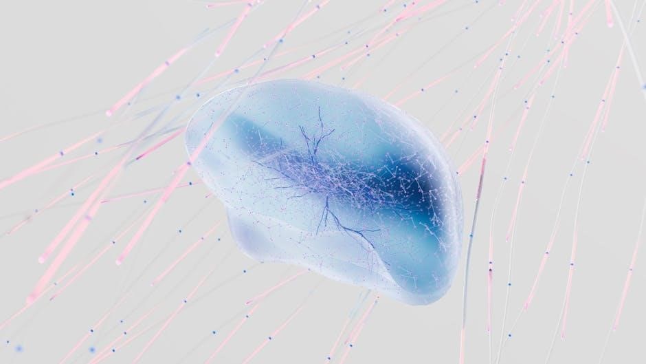
Brain anatomy PDF resources extensively detail the cellular building blocks of the nervous system, focusing on neurons as the fundamental functional units and glial cells as vital support structures. These documents, often adapted from comprehensive anatomy texts, present diagrams illustrating neuron structure – dendrites, axons, and synapses – and their role in electrochemical signaling.
They differentiate between various glial cell types, including oligodendrocytes, which, unlike Schwann cells in the periphery, form parts of multiple neurons. PDFs explain how glial cells provide structural support, insulation (myelin), and nutrient transport.
Clinical notes within these resources connect cellular dysfunction to neurological disorders. They emphasize the importance of understanding these microscopic components within the context of the larger 1.3-1.5 Kg human brain, providing a foundation for comprehending complex neurological processes.
A. Neurons: The Basic Functional Units

Brain anatomy PDF guides dedicate significant sections to neurons, defining them as the core functional units of the nervous system. These resources illustrate neuron structure – dendrites receiving signals, a cell body integrating information, and an axon transmitting impulses – often with detailed diagrams.
They explain how neurons convert stimuli into electrochemical signals, crucial for all brain functions. PDFs detail synaptic transmission, the process by which neurons communicate at junctions, and the role of neurotransmitters.
Clinical correlations within these documents link neuronal dysfunction to various neurological conditions. They emphasize the importance of understanding neuronal anatomy for diagnosing and treating brain disorders, within the context of the larger brain’s complex network. These resources often highlight the neuron’s role in the 1.3-1.5 Kg adult human brain.
B. Glial Cells: Supporting the Neurons
Brain anatomy PDF resources extensively cover glial cells, emphasizing their vital supporting role to neurons. These documents detail various glial cell types – astrocytes, oligodendrocytes, microglia, and Schwann cells – and their specific functions.
PDF guides illustrate how oligodendrocytes, unlike Schwann cells in the periphery, form parts of multiple neuronal myelin sheaths, enhancing signal transmission speed. They explain astrocytes’ role in maintaining the blood-brain barrier and providing nutrients to neurons.
Microglia’s immune function within the brain is also highlighted, alongside the structural support provided by these cells. Clinical sections within these diagram-rich resources connect glial cell dysfunction to neurological diseases. Understanding glial cell anatomy is presented as crucial for comprehending the overall health and function of the 1.3-1.5 Kg human brain.
VII. Brain Anatomy Diagrams and Resources (PDF Focus)
Numerous brain anatomy PDF resources are available, offering detailed diagrams and comprehensive explanations of the central nervous system. These materials, often adapted from textbooks like Human Anatomy & Physiology by Marieb and Hoehn, are invaluable for students and medical professionals.
PDF documents frequently include labeled illustrations of the cerebrum, brainstem, cerebellum, and deep brain structures, facilitating accurate identification of key parts. They often present both gross anatomical views and cellular-level depictions of neurons and glial cells.
Resources emphasize the importance of understanding the embryonic scheme of brain development. Many PDFs provide interactive exercises for labeling brain structures and assessing comprehension. These resources aid in visualizing the complex organization of the adult human brain, weighing 1.3-1.5 Kg, and its functional divisions.
VIII. Clinical Relevance: Understanding Brain Anatomy for Diagnosis
A robust understanding of brain anatomy, often reinforced through detailed PDF resources and diagrams, is crucial for accurate medical diagnosis. Identifying specific brain parts affected by injury or disease relies heavily on anatomical knowledge.
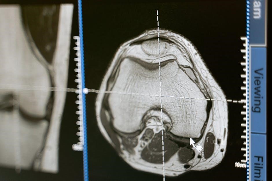
PDF materials detailing the nervous system’s divisions – central and peripheral – aid in localizing neurological deficits. Knowledge of structures like the cerebellum, corpus callosum, and frontal lobe is essential for interpreting imaging scans and neurological examinations.
Clinical applications range from diagnosing stroke and traumatic brain injury to identifying the neurological basis of behavioral changes. Resources adapted from texts like Marieb and Hoehn’s Human Anatomy & Physiology provide a foundational understanding. Accurate anatomical assessment, considering the brain’s 1.3-1.5 Kg weight, is paramount for effective patient care and treatment planning.